
Understanding Macular Degeneration: What You Need to Know
"My mother has been told she has macular degeneration. Does that mean I'm going to get it too?"
It's one of the most common questions we hear from patients in their fifties and sixties—and it's one worth taking seriously.
Age-related macular degeneration (AMD) is a leading cause of sight loss in people over 50 in the UK. It affects hundreds of thousands of people nationwide.
One challenge with AMD is that it often develops gradually. In many people, the early stages produce few or no noticeable symptoms, meaning changes may be present before vision is affected. That window — when AMD may be detectable but has not yet caused noticeable visual symptoms — is precisely when regular eye examinations and retinal imaging can be particularly valuable.
WHAT IS THE MACULA — AND WHAT DOES IT DO?
The macula is a small but extraordinarily important area at the centre of the retina — the light-sensitive layer at the back of the eye. It provides your central, detailed vision: reading, recognising faces, watching television, driving, and threading a needle.
AMD does not cause total blindness — peripheral vision is usually preserved. But losing central vision can be profound. Reading a book, seeing the faces of people you love, recognising someone across a room — these become increasingly difficult as the condition progresses.
That is why early detection isn't simply a clinical nicety. It can make a significant difference in preserving vision and maintaining quality of life.
DRY AMD VS WET AMD: THE KEY DIFFERENCE
AMD has two types, and understanding the difference matters.
Dry AMD (85–90% of cases)
Dry AMD is caused by drusen — fatty deposits — accumulating beneath the macula.
Progression is usually slow and gradual over many years. No treatment currently reverses dry AMD, so monitoring and lifestyle measures matter.
Wet AMD (10–15% of cases)
Wet AMD is caused by abnormal blood vessels growing beneath the retina and leaking fluid.
Although less common than dry AMD, it can progress rapidly and requires urgent assessment and treatment. Anti-VEGF injections can often stabilise vision and, in some cases, improve outcomes when treatment starts promptly.
The critical point: OCT retinal scanning can often detect signs of AMD before noticeable symptoms develop, helping us monitor changes and advise on appropriate next steps.
EARLY WARNING SIGNS — AND WHY MOST PEOPLE MISS THEM
Dry AMD in its early stages often produces few or no symptoms. As it progresses, the first signs are typically subtle:
Blurring or haziness in the very centre of vision
Colours appearing slightly washed out or less vivid
A need for more light than before when reading
Straight lines appearing slightly wavy or bowed (called metamorphopsia — an important warning sign)
A blank or blurred spot in the centre of vision in more advanced cases
WHEN TO CALL US URGENTLY
If you notice any sudden change in your central vision—new distortion, a blank spot, or straight lines that have become wavy—please contact us the same day.
For wet AMD in particular, early assessment and treatment offer the best chance of preserving vision. Do not wait to see if it improves.
WHO IS AT RISK?
Risk factors you cannot change.
Age — AMD is uncommon before the age of 50 and becomes increasingly common as we get older
Family history — having a close relative with AMD can significantly increase your risk of developing the condition
Genetics — certain inherited genetic factors are associated with an increased risk of AMD
Sex — AMD is slightly more common in women, partly because women tend to live longer
Risk factors you can influence
Smoking — smokers are up to four times more likely to develop AMD. The risk persists for years after stopping
UV exposure — wear UV400 sunglasses outdoors, particularly near water, sand, or snow
Diet — a diet rich in leafy greens, oily fish, and antioxidant vitamins is associated with reduced AMD risk
Cardiovascular health — high blood pressure, high cholesterol, and obesity are all associated with elevated AMD risk
HOW WE DETECT AMD AT ARTHUR HAYES EYECARE
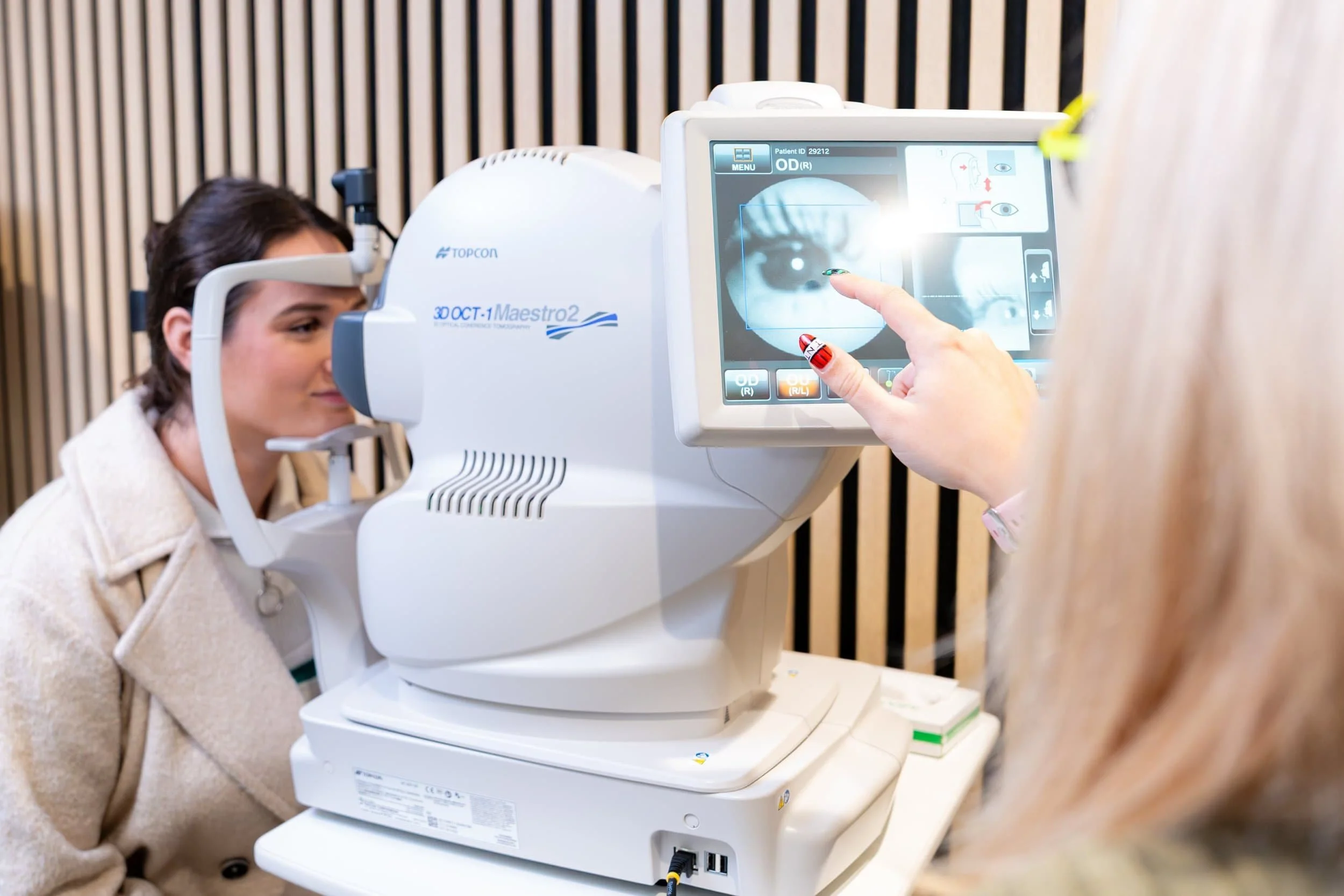
We use the Topcon OCT Maestro2 — one of the most advanced retinal imaging systems available in independent practice.
OCT (Optical Coherence Tomography) produces a detailed cross-sectional image of the retinal layers, comparable in principle to an ultrasound but using light rather than sound.
OCT can detect:
Drusen beneath the macula before they affect vision
Sub-retinal fluid associated with wet AMD
Geographic atrophy — areas of retinal cell loss in advanced dry AMD, measurable over time
OCT imaging takes less than a minute, requires no eye drops, and involves no contact with the eye.
If you have a family history of AMD, or if you are over 50 and haven't had retinal imaging as part of a recent eye exam, it may be worth discussing OCT scanning at your next appointment—even if your vision currently seems normal.
NUTRITION AND AMD: WHAT THE EVIDENCE SHOWS
Lutein and Zeaxanthin - These carotenoid pigments are found in kale, spinach, broccoli and egg yolks.
Higher dietary levels are associated with a reduced risk of advanced AMD.
Omega-3 Fatty Acids - Found in oily fish such as salmon, mackerel, sardines and herring. Two portions of oily fish per week are associated with a modest reduction in AMD risk.
AREDS2 Supplements - The Age-Related Eye Disease Study 2 found that a specific combination of antioxidant vitamins and minerals — 500mg vitamin C, 400 IU vitamin E, 10mg lutein, 2mg zeaxanthin, 80mg zinc, and 2mg copper — may help reduce the risk of progression to advanced AMD in suitable patients.
These supplements are recommended for certain patients with intermediate or advanced AMD, not as a general preventive supplement. We will advise whether supplementation is appropriate based on your individual findings.
IF AMD IS DETECTED: WHAT HAPPENS NEXT?
Dry AMD - We will recommend a monitoring schedule — typically every six to twelve months — and discuss lifestyle and nutritional measures. No treatment currently reverses established dry AMD, so the primary goals are preserving vision and monitoring progression.
Wet AMD - If OCT reveals fluid beneath the retina, we will refer you urgently to the NHS medical retina service. Anti-VEGF injections are highly effective when started promptly, and the sooner treatment begins, the better the potential outcome.
Low Vision Support - For patients with more advanced AMD, our low vision service provides practical support, including:
Magnification aids
Lighting advice
Referral to specialist rehabilitation services
Reduced central vision does not have to mean an end to reading and independence.
BOOK YOUR OCT RETINAL SCAN
If you are:
Over 50
Have a family history of AMD
Have not had a full eye examination, including retinal imaging, in the past year
Please consider booking an appointment at Arthur Hayes Eyecare, Crowborough. An OCT retinal scan takes less than a minute and may help identify changes that could affect your future vision.
Disclaimer: This article is intended for general information purposes only and does not constitute medical advice. If you have concerns about your eye health, please book an appointment with a qualified optometrist.
Arthur Hayes Eyecare is registered with the General Optical Council.